What is an Arteriovenous Malformation (AVM)?
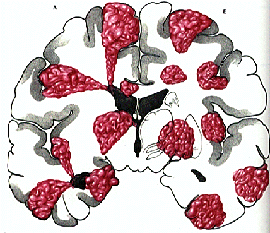
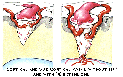
This patient presented with sharp head pains and loss of consciousness as a result of a seizure. He is a 47 year old Asian-American in relatively good health. After a thorough history, the patient was taken for CT. Upon analysis of the scan, it was found that this patient had “blood one the brain.” After further analysis with MRI, it was determined that the reason for this hemorrhage was AVM. AVM is short for Arteriovenous Malformation and are extremely rare. There are only about 250,000 reported cases. AVMs are abnormal collections of blood vessels in which arteries lead directly into veins, nidus. These blood vessels don’t follow the normal network path using the smaller blood vessels such as arterioles and venuoles. In turn, pressure develops on the arterial end of the vessel causing hemorrhage and swelling. This condition, normally present from birth, presents the most danger from 10-55. If no problems have developed by this time, the chances of problems appearing decrease rapidly. On the other hand, if there has been a hemorrhage during this time, then the chances for more and more intense hemorrhages increases rapidly.
There are at least four different types of treatments for AVMs. Radiation, angiography, surgery and no treatment are all valid choices given the particular circumstances of the AVM. The size, position and complexity of the blood vessel arrangement are all things that must be taken into account.
Now that the requisite background has been given, let’s jump into the amazing case. Based on the factors from the previous paragraph, it was the determined that the best course of action for this particular patient was embolization.
What is Embolization Therapy?
“Embolization is a method of plugging the blood vessels of the AVM. Under X-ray guidance, a small tube, a catheter is guided from the femoral artery in the leg up into the area to be treated.
A neurological exam is performed before and after a small amount of medicine is injected. This can help tell if the vessel that feeds the AVM also feeds normal and important portions of the brain. After this, a permanent agent is injected into the AVM and the catheter removed. This is repeated for each vessel that feed the AVM.”
Excerpt from http://www.aplasticcentral.com/avm/
So, after getting the patient on the table for the procedure, things went extremely well. The main arteries feeding the AVM were isolated and epoxy or glue was used to close the vessel. This procedure proved be very complex as the angiogram show more vessels than were revealed by MRI. With the information from angiography, a 3D image is generated that obviously gives the physicians a more complete and detailed image. At this point, the game plan changed. Dr. Riina decided to plug as many arteries as possible but the craniotomy would be necessary to give the patient the best chances of a full recovery. Because of the size of the hemorrhage, the patient was scheduled for a craniotomy the next morning.
Briefly, the procedure for attacking an AVM is to clip all the blood vessels that feed the AVM. After clipping, imaging and visual inspection allow the physician to excise the AVM in its entirety. This procedure is much like that of removing a tumor. It is imperative to close off all vessels that feed the abnormal mass or the result can be severe hemorrhaging and ultimately loss of life. Fortunately, Dr. Riina was able to perform the procedure without any complications. The patient is expected to make a full recovery although there will be some discomfort and maybe minimal memory loss.
For your enjoyment, I have enclosed a links to diagrams of an AVM and a movie of an AVM removal via craniotomy, which has two parts.

http://www.brain-surgery.com/avm.gif

http://www.brain-surgery.com/avm2.gif
http://www.brain-surgery.com/avmpt1.avi
http://www.brain-surgery.com/AVMPRT2.AVI

0 Comments:
Post a Comment
<< Home