Tuesday, August 08, 2006

My Farewell

Well, now its time to say, “Farewell.” Overall, I have had an awesome, unforgettable experience. I would like to thank God for giving me everything that He has to sustain me thus far. This is only possible because of you. I would like to thank Dr. Howard Riina in Neurological Surgery. Dr. Riina, you made this an enjoyable experience. You taught and showed me a great deal in these 6 weeks. Because of it, I have a new appreciation for medicine, especially neurosurgery. Next in line is Dr. Kyle Chapple, a Neurological Surgery fellow. I would like to thank you for my interaction with you and your honesty. You also taught me volumes of information and gave me valuable perspective from that of a resident and fellow. Beyond that, you made the experience fun and enjoyable. To the staff in the Neurological Surgery office and in Interventional Neuroradiology, my hat is off to you all. I felt very comfortable with you all and thank you for your time and insight given to me during the procedures. I would like to also thank the Biomedical Engineering Department of Cornell University and all pertinent faculty and staff that made the 2006 Summer Immersion possible. Lastly but not least, I would like to thank my beautiful wife, Dr. LaShanda T. J. Korley, and my unborn child due August 31. You could have objected but you supported me throughout. I can’t wait to see you. To Baby Clara, I am glad that you waited on your dear old dad. We talked several times about you waiting to arrive after the program in NYC was finished. You are listening well at any early age and I truly appreciate it.

Monday, August 07, 2006

Acknowledgments and Thank You

I would like to acknowledge the following people who helped make my experience at the Cornell Medical School and NYPH one that I will never forget:

Dr. Jonathan W. Weinsaft
Dr. Matthew Janik
Dr. Matthew D. Cham
Dr. Howard Riina (for allowing me to observe neurosurgery)
Dr. Karl Krieger (for letting Julius and I observe the CABG)
Dr. Jorge R. Kizer (for being a test subject for your THICK study)
Nurse Lily S. Yee (for explaining the echocardiogram I received for the THICK study)

Thank you to all the nurses and residents in 4 North for answering all my questions; to the technicians in the MRI, CT, SPECT (aka nuclear medicine), Echocardiogram, Interventional Radiology (neurology), and Catheterization (cardiology).

Thank you to my fellow colleagues in the program: Julius for allowing me to see how that aneurysm was clipped along with our adventures throughout the city, Rachel for being able to organize all the events and outings, Christine and Flor for the games of pool, Vishal and Ben for the card and domino games, Jun and Jesse for heading out to grab wings and beer, Evan and Alex for the best locations in the Wholesale district, Robbie and Daniel for your experiences in HSS, and to everyone who related their stories and projects during our meetings and experiences.

A big thank you to Dr. Yi Wang for organizing this opportunity to experience the medical field and also to Belinda Floyd for taking care of all the paperwork, all the arrangements, and accommodations (this would not have worked out if it were not for you!).

Thank you once again to everyone. This experience will not be forgotten.

Week Six and Its Over

Its amazing to see how fast six weeks in NYC can fly by…it seems that it was only a few days ago that we had arrived at the medical school and were trying to figure out way about the hospital and now we’re all packing up and getting ready to head back home to Ithaca with the research that awaits our return.

This week began in the OR where Julius and I were able to observe a double bypass open heart surgery, otherwise known as a CABG (coronary artery bypass graft). The patient was a 72 year male that needed this surgery to regain circulation in other parts of his heart due to occlusions in the main arteries that supplied blood. The patient was put under anesthesia and was prepped for the operation with a cardiothoracic fellow working on opening up the chest cavity and another surgeon that was harvesting two vessels from the patient’s legs. The surgeon performing the vessel harvest made two incisions on the leg and arthroscopically inserted a tool that had a fiber optic camera, a pair of cauterizing scissors, and hook that allowed the surgeon to capture and isolate the vessel of interest. It was amazing to see the speed and accuracy that the surgeon worked at and in a matter of minutes had isolated the vessel, cut off all the branching arteries from it, and had it outside of the patient and was cleaning the vessel up. During the clean up process, the surgeon attaches little staples to where the cauterized branching vessels were (for this reason during an angiogram the staples appear on the x-ray) and then attaches the vessel to a syringe filled with saline and pressurizes the vessel to determine if there are any leaks. Once the vessel is cleaned up, its placed in a holding container with saline and then the surgeon proceeds to work on the other leg and to retrieve another vessel.

During this same time, the cardiothoracic fellow has been working on the patient’s chest cavity and has cracked the rib cage (actually sawed through the sternum with a power saw and then opened it up with a chest cavity spreader). He then proceeded to cauterize all the blood vessels that he had cut through and began to remove the pericardium that covers the heart. As soon as he had the heart uncovered, he then proceeded to get ready for the bypass which involved inserting hoses into the chest cavity and hooking the heart up to a heart and lung machine. This was an amazing sight to see the amount of blood and saline that is used during the recirculation process and the color and thickness that blood takes on when it is flowing in such volumes. After the patient was hooked up to the machine, the heart was chilled to 10C and the chief surgeon Dr. Krieger came into the OR. Even at 10C, the patients heart was strong enough to keep beating and the surgeons then set on with the second part of the procedure which was identifying where the new vessels were going to be grafted on to the heart. During this time, Julius and I took turns to stand by the anesthesiologists station and look down onto the heart and what was going on. After the back vessels had been attached to the patients heart, Dr. Krieger looked up and asked what we were doing in the hospital, our reply was that we were graduate students in the Immersion program and we were observing. He replied, “Observing? You’re not observing. Get up here and see what we’re doing.” Now this was an amazing treat, we were able to stand again right by the anesthesiologists station and look down on the front part of the heart as it continued to beat. During this part of the procedure the surgeons took the aorta and clamped it, and then punched holes into it to attach the vessel grafts that were sewn onto the punched holes. While this was going on, Dr. Krieger was talking to us and describing the whole procedure in all its details. The patient was taken off the heart and lung machine afterwards without needing his heart restarted because it had continued to beat during the whole procedure and no bleeding was evident. The fellow then closed up the patients chest and the procedure was over in under 6 hrs. To say the least, that procedure was just amazing.

The rest of the week was spent trying to got old MRI records of patients restored on the workstation so that I could begin tracings for my project with Dr. Weinsaft, but that did not happen till Wednesday. I managed to complete a few tracings before I left, but ended up having to leave the rest of the project for Dr. Janik (a fellow scientist who had been working with Dr. Weinsaft on a previous collaboration). In addition to working on the project, I was able to sit in on a few more MRI/CT readings and go in for a few more morning rounds with the residents on 4 North.

Wednesday evening we had our wrap up meeting with Dr. Yi Wang and had a great meal at an excellent sushi place (except for the air conditioning breaking down), while on Friday it was one last trip to the 55th Street MRI building to see a few more readings, followed by a quick lunch with Dr. Weinsaft, and then last minute packing and laundry before the bus left back to Ithaca.

Overall I must say that the experience this summer was an amazing one. Getting to work at the medical hospital allows us engineers to see how the medical system works, how the doctor and patient interactions occur, how current techniques are used to treat and diagnose disease, and how we can use our own skills to help improve specific problems that currently exist in the medical field. This opportunity that is afforded by Cornell should be extended and modeled at other schools that have a Biomedical program because through these programs, the students really begin to see what the fields really need in terms of devices and new inventions that can bridge the biological and engineering gaps that exist.

Friday, August 04, 2006

Oh, What a Feeling!

This week offered new experiences. We were finally able to “coil” a patient that had previously been rescheduled due to complications. I am glad that it happened this soon as the patient had an aneurysm measuring 14 x 11 mm. Luckily, there was no hemorrhage or any other complications. The case went very well and the patient is expected to make a full recovery.

Cardiothoracic surgery is amazing. I was afforded the opportunity, along with my colleague, Jan Kostecki, to observe a CABG (coronary artery bypass graft). In this procedure, the patient’s vasculature around the heart was badly compromised. The plan was to endoscopically take veins from this patient’s legs to replace the vasculature of the heart. The process of removing the veins from the legs of the patient has come a long way. In the past, a surgeon excised the vein by scalpel, leaving horrible scars and a substantial would to heal from. This procedure was done with only to small incisions above and below the inside of both knees. It was very exciting to see how technology had changed.

While a doctor was getting the replacement vessels, another surgeon was working to prepare the thoracic cavity. For the procedure, the chest was opened via a scalpel and sternum saw. The appropriate vessels were cauterized to minimize bleeding and the patient was put on the heart and lung bypass machine. Cardiothoracic surgery is unlike neurosurgery where a screen shows you everything that the surgeon is doing. In cardio surgeries, one needs to be very close to see what is going on. Both Jan and I were constantly trying to walking around the room to get the best spot. At some point, one of the nurses told us that we could stand on a step stool behind the patient’s head so that we could have a better perspective. We were cautioned to only stay there momentarily as not to upset the attending surgeon, Dr. Krieger. By the time Dr. Krieger showed up, we were only on the outskirts, making sure not to be in anyone’s way. He saw us looking on and asked who we were. I explained that we were graduate students in BME from the main campus. At that point, he told us to reclaim our prior position near the patient’s head. He began explaining everything. The anesthesiologist had to ask us to move to check on the patient’s vitals. Those moments were very short as Dr. Krieger was intent to explain the finer points of this procedure and the procedure was progressing well. We were blown away by the procedure and his kindness. We learned so much and it was really exciting to see a person’s heart and lungs. Books can’t really capture what my eyes beheld. The heart’s beating was fascinating. Even when cooled with ice, this person heart was so strong that it continued to beat. After finishing the procedure, the person was immediately able to come off of the bypass machine and use his own heart to pump blood to his entire body. It was amazing to see and very fortunate for this patient. The procedure was a success and cardiothoracic surgery is a very cool place to be.

My experience really can’t be described in words. During these 6 weeks, I have tried to capture the highlights but my words pale in comparison to the actual experience. I hope that you all have enjoyed the depictions of my experiences. I hope that in the future I am afforded this opportunity again. Besides, the procedures, I was able to have meaningful dialogue with some physicians about potential collaborations and consulting opportunities after graduation. I am very excited and look forward to the future as I think that it will be a bright one. Also, I have a new appreciation for the hard work that so many put forth to take care of patients and give them the best opportunity for a successful recovery. I am glad to have been a participant in the 2006 Biomedical Engineering Summer Immersion at Weil Medical College of Cornell University.

Tuesday, August 01, 2006

5th Week Brings A Change to the Routine

So Monday and Tuesday were the normal routine where I got up early, went in for rounds with the residents on 4 North at 8am, and observed cases till about noon. After that was done, I headed down either to the MRI room or the SPECT room or the CT room and read images with the fellows and attending doctors that were in the room. It has become quite easy to diagnose the common diseases that the doctors see on a regular basis in the hospital and I have become proficient at using the software and manipulating the data/images to make a diagnosis or to show a person that is observing where the problem lies.

During these two days as well, I cleaned up my patient data set that I had already complied and excluded some patients that had complicating factors or that there was not enough data present to make sure that they fit our study. After going through this group and making sure that the patients that we had all fit our criteria, I took the list down to the MRI room to have the data retrieved and placed on the workstation so that I could begin the real data analysis. Suffice to say, retrieving the old records is always put on the back burner, and as I write this blog a week later, I am still waiting for the patient files.

In addition to the work that I had done, on Friday I met up with Dr. Matthew Janik and he showed me the correct way to trace images on the MRI software (the tracings use a complex algorithm that allows for the measurement of size, shape, and functionality of the heart). These tracings are the method in which I am going to obtain the data for our study, but one thing that is blatantly apparent is that the tracings are all subjective to the person that is doing the tracing. Since an MRI image takes a slice of the object, the image has items that are both in and out of the plane, and these items can be either traced or not traced. Therefore, the tracings of the images that we have will be evaluated by three experienced MRI imaging experts and then those opinions will be taken into account for the final values obtained.

Now the fun part of the week begins…well Wednesday and Thursday. So my clinician left for a conference and since I could not do anything related to my project I talked with Julius and asked if I could observe some of the medical procedures that had been going on in his area of expertise. On Wednesday I was able to see an aneurysm get clipped in a 30 year old woman. When we walked into the OR, the woman was already on the table and the fellow had already removed her scalp, pinned it in place, and was beginning to clean up the skull so that there could be access to the brain. At the same time, there was an incision made in the woman’s neck and a clamp was placed on her carotid artery to stop blood flow into the region where the aneurysm was. The first thing that struck me in comparison to the imaging that I had been doing these last few week was the smell of the OR. The smell of tissue being cauterized is something that many people don’t forget, and for those that wonder what its like, I think its like when you singe the hairs on your arms when reaching over a stove or a camp fire. The smell is really that pungent, but you begin to be desensitized to it quite quickly. After that I also realized the amount of instruments and technology that goes into a surgery and how everyone has a specific role to play. Once the skull was cleaned off the surgeon took a tool that looked like a Dremel and went to work cutting out a portion of the skull where the aneurysm was located. After removing the skull and dissecting the way down, the aneurysm was pinpointed on the microscope and tv monitors that were present in the room. The next step was to take small clips (yes, clips like those used to hold laundry on the line) and clip the blood flow to the aneurysm. To make sure that all the blood flow was stopped, both ultrasound was used to see if there was any flow through the area and a fluorescent dye was injected that could be seen under infra red monitors. The clip was in the correct place and then the surgeon popped the vessel and closed up the region. That was an amazing procedure and took about 5 hrs to complete.

The rest of Wednesday and Thursday were spent in the Interventional Radiology room on the 5th floor where I was able to observe catheterization procedures that mapped the blood flow in the brain. These procedures were the same as those as I had seen in the cardiology catheterization room, but the focus was on mapping flow through the brain. One patient that we had seen had a tumor that was detected by CT, but upon looking at the blood flow via that catheterization the decision was not to perform surgery because there was no single vessel feeing the tumor and there was no risk to the patient. The rest of the cases were also uneventful but it was interesting to see how the same procedure could be used to diagnose other conditions in the body.

Sunday, July 30, 2006

Are We There Yet?

Most of this week was spent trying to define the parameters of my project. I am very lucky to be shadowing Dr. Riina as he is a very busy surgeon. However, that also means that there is less time for defining project parameters and such. However, at this stage my project proposal looks as such.

Before any procedure or operation, most patients desire to know their chances of a successful outcome. In many situations, patients will not move forward with the proposed procedure if their chances are not favorable. Obviously, no doctor keeps track of every single case so it is very hard to calculate any success rate associated with any given procedure. Even more confounding are the different variables associated with each case. Ultimately, a physician’s attempt to give accurate prognostic information is a calculated guess. One can be certain that by no means does a physician intentionally mislead a patient as they are not computers and certainly not omnipotent. What if it was possible to devise a scale with predictive values based on a physician’s body of work? This prognostic indicator would be semi-quantitative and give a more accurate assessment of the patient’s chance of having a successful procedure. It is proposed that the scale would consider and account for the multiple variables effecting patient’s with an aneurysm such as location, size, age of patient. The value generated would then correlate to percentage ranges to be determined. For this type of study, a sample of historical data would be taken from multiple cases. After analysis, this information would be used to incorporate the multiple variables existing in aneurysm cases to be used as tool to give patients a truer assessment of their relative chances of a successful procedure.

This week was slow. Dr. Riina was in the hospital for two days so on the off days, I observed Dr. Pierre Gobin, a neurology interventional radiologist. He performs many of the same procedures as Dr. Riina. I saw an interesting case where this child had a retinoblastoma. To treat this, Dr. Gobin embolized the tumor by stopping its supply of blood with polyvinyl alcohol particles. At this stage, it is a possibility that the tumor will be carefully excised by surgeons at Memorial Sloan Kettering Cancer Center.

I also had a chance to get back to the OR. I saw another aneurysm clipping. This was much like others I have described but they never get old. A couple of the really cool things about this particular procedure were the location of the aneurysm and the clipping of the lower vessel that fed into the aneurysm. The aneurysm was located near the optic nerve. Because of the complexity of the area, a temporary clip was placed on the external carotid artery to stop blood flow to the aneurysm. At this point, Dr. Riina proceeded to clip the aneurysm. For this case, two clips measuring more than 10 mm each were used. One was initially in place but was not sufficient to stop blood flow to the aneurysm. This was because of the location and neck size of the aneurysm. After both clips were in place, Dr. Riina used infrared imaging and a Doppler instrument to check the blood flow integrity and assure that the abnormal vessel was properly repaired.

Well, the last week of this immersion is approaching quickly. I am looking forward to making more progress on my project. I will be collecting the much needed patient data to get the study underway. After receiving the patient data, the meat of the project will begin as I need to investigate if a correlation exists between a successful procedure and any variable or variables associated with each patient’s particular procedure. If all works well, a recipe may exist for each patient to have a successful procedure. Maybe as important, this study could offer semi-quantitative prognostic information for Dr. Riina’s patients in the future.

Monday, July 24, 2006

Jan Kostecki – Immersion Student but Test Subject Too

So this week started off a little differently than the others have; instead of going to 4 North for 8am rounds, I began the day by going to the 2nd floor GCRC to get a full blood work up. You may be wondering why I needed to get a workup, but this was for Dr. Kizer and his THrombophilia In Cryptogenic stroke (THICK) study. I was recruited by Lily, who is a echocardiology nurse on the 4th floor. The study started off with filling out the standard medical history questionnaire, followed by a round of questions about my background, and a bunch of other paper work that needed to get done. After that I was lead into a room where a nurse was going to take the blood, at first I thought this was going to be like any other blood draw, but it turns out that they needed 15 vials of blood, each vial was only a tablespoon in size, but still 15! I’m normally ok with having my blood drawn, but I’ve never had the need for an IV needle, so when the nurse put the IV in and even before she began to take the blood, I had begun to see stars and soon I couldn’t see at all. This followed by a cold sweat, becoming completely pale, and on the verge of passing out. This whole time I was talking to the nurses and had this big grin across my face, and they thought I was just messing around with them. As soon as they saw me turn white and break out in a sweat they new I was not kidding. I stayed conscious through the whole draw, and regained my vision to see the last five vials get filled up. At this time, the whole room was filled with nurses just talking about what had gone one, suffice to say, I had put on quite a spectacle for the floor. After getting some juice and taking a few minutes to regain my composure, I went up to the fourth floor and by this time news had already spread to Lily who had a good chuckle. I then went in to get my echocardiogram and Lily took her time to show and describe the different parts of my heart, what she was looking at, along with the anatomy of the heart and blood flow. After the basic imaging of my heart was done, Dr. Kizer came in and took saline that had been oxygenated and injected it into the IV that was in my right arm. Upon injection of the saline, the echocardiograms revealed the oxygenated bubbles in the saline as they traveled to my lungs and were then expelled (no need to worry about a bubble of air heading to my brain). The study was to see if there was a hole between the two atria of my heart, and subsequently revealed that I was a normal control patient. The group is still looking for another 300 control subjects, and if you are interested please let me know and I will get you the contact information. A plus side is that the blood work and the echocardiogram are all free, so you’re looking at around $4000 for a whole cardiac work up for free!

After having that fun of a morning, I read MRI images with my clinician (mainly normal hearts so that I could see what a normal heart looks in comparison to a diseased one), and then was lead into the EP lab where I observed the mapping of a patients heart. The patient had passed out a few times and the doctors wanted to know if it was due to a electrical conduction problem (either the heart was beating too slow or too fast). The room looked like a standard catheterization lab where I had observed angiograms, with a slight modification. There was a big black box in the control room and two computer monitors that had about 30 different electrical signals being displayed. Here I was able to observe how the doctors were able to map the electrical signals across the heart and diagnose where the problems were occurring. It turned out that the patients electrical system was function correctly and did not have any abnormalities.

Tuesday through Friday were back to the standard routine of getting up for 8am rounds on 4 North, followed by meeting up with Dr. Weinsaft and reading images of MRI, CT, and nuclear exams. I also attended a seminar for fellows at the hospital on how to read nuclear exams (specifically related to SPECT images). From what I had already learned first hand with Dr. Weinsaft, this was basically a review course for me and I could already pick out the problems in the heart before the presenter got to them.

In addition to reading images through the week, I continued to work on my project. Since I had the list of patients that we would be using for the study, I now had to compile a demographics sheet that had the patients age, previous medical history, family history, and list of medications that they were on before getting their MRI and echocardiograms done. Many of the patient’s records were easy to access and the database entry was straightforward. There was however a handful of patients that did not have their records on file, so I learned how to use the hospitals other database to locate the patients records. In these records, I had to go through them page by page to see what the physicians had listed. This was more time consuming because I had to decipher hand writing of physicians, and what they do say about a doctors hand writing is true: its completely illegible most of the time, but then there are those exception to the rule which makes it so much easier to find the information that you need.

Overall it was quite a eventful and fun week, and I learned many more new things, not only about the medical field, but also about my own physiology.

Listed below are a few pictures of my mentor, Dr. Jonathan Weinsaft and I reading CT images. Basically my days are spent in small room looking at computer monitors.

The God Complex: Truth or Myth

This past week has been very enlightening. From my perspective before this experience, it seemed that surgeons were mavericks blazing a trail into a new horizon. TV made surgeons out to be egotistical maniacs that had the “God Complex.” As my time has progressed here, I have slowly learned of the teamwork needed between all of the people in healthcare to effectively treat a patient. Many patients have multiples issues and it is only with good teamwork by way of great communication that these patients have their best chance of successful treatment. In my experience here at Weill, I have not found a surgeon that could be accused of having a “God Complex.” It is easy to understand why they need so much confidence. As a resident and or fellow, they are constantly torn down by the attending physicians. Yes, it is deliberate but the intentions are not malicious in nature. One of the goals is to create people of strong character who will stand up for what they feel is the best treatment for a patient. Surgery has proven to be a field where aspirants should have a “thick” skin. The surgeons that I have met have been very confident. I am sure that I only want surgeons operating on me that have confidence. I think that there is a fine line between overconfidence and confidence; the surgeons here tread carefully.

Last week proved to be very challenging. There was more of the same but some cases draw a person in more than others. I asked a Neurological Surgery fellow,”How do you not get emotionally attached?” He began telling me a story about his third year in medical school. As he entered an examination room to see a patient, the patient took a deep breath and died before his very eyes. He explained that becoming emotionally vested in the patients that he cared for would be too taxing. Before coming for this Summer Immersion program, I probably would have thought that to be harsh and the easy way out. Now I understand how true his statements are. Each week there has been at least one case in which I felt some attachment. Sometimes your heart can not help caring for patients with compelling circumstances. In my position, shadowing a clinician, I began feeling the weight of this burden. It seems an almost impossible task to carry all of that baggage around. I began thinking about the patients too often. I was vested in their successful recovery. Being vested in their recovery alone would probably be ok, but that’s not life. Recovery is not always the path chosen. Death occurs often and sometimes without warning. I can’t begin to imagine the bag of bricks that I would have to drag around if I became attached the patients that we have seen thus far. Well, enough of that, we are moving on to the fun stuff!

A 36 year old woman was brought to the ER after suffering from numbness on her left side. We were told by her husband that her chief complaint before loosing consciousness was severe headache and splitting pains. For whatever reasons, she thought this would pass but it obviously ended up being more than she bargained for. While in the ER, she had a seizure involving her torso and left leg. This seizing began spontaneously. A CT scan showed an area of hypodensity measuring 3.3 cm with associated hemorrhage. At this stage, it was thought that the patient could be suffering from an AVM. On day 2, a second seizure was observed and treated. The patient’s movement was severely retarded. The patient was transferred to the NYH NSICU. After angiography, it was determined that this patient was suffering from sinus thrombosis. In this condition, a clot, thrombosis, has migrated the vasculature of the head and become trapped in one of the sinuses, venous system for the head. In short, blood was collecting in the patient’s head causing damage to surrounding tissues which resulted in paralysis on the left side.

After review angiography footage, a team of physicians were called in to consult on this case. It was determined that the best course of action would be to wait on TPA (tissue-plasminogen activator) treatment as the patient’s movement on her right side had become better. TPA involved certain risks and it seemed that the IV medications were helping. One of the risks was bleeding around the clot that had formed in the sinus. If this bleeding was unable to be controlled, a vegetative state could be the result. On the other hand, TPA could dissolve some or the entire clot and restore drainage. After no improvement over 12 hours, a couple of rounds of TPA were administered in the NSICU. The patient has improved at his point but is still undergoing treatment. She is a soldier with much to fight for as she is a wife and the mother of 4.

More on TPA http://www.americanheart.org/presenter.jhtml?identifier=4751

Thursday, July 20, 2006

Week #3: Opposite ends of the spectrum

Another week in the trenches and a whole new set of cases that have been read in MRI. I think that my experience with Dr. Prince has been a bit different from everyone else's because my focus is on MRI, not speaking to patients and watching surgeries, but that doesn't make it any less interesting!

We're all engineers or at least aspiring to be engineers and MRI is a great example of how scientists and engineers can reshape the medical field. It can all be traced back to Raymond Damadian's original patent filed in 1971 and issued in 1974 (Pat. No. 3789832). Damadian's patent and several journal articles disclosed that he had discovered certain mouse tumors displayed elevated relaxation times compared to normal tissues. This Damadian hypothesized could be used in humans to identify diseased tissue from healthy tissue with much greater contrast than offered by X-ray and Ultrasound. This discovery combined with recent work in cryogenics made possible the construction of large super-conducting magnets necessary for MR-imaging in humans and thus MRI was born.

The story goes that Damadian and colleagues designed and built their own whole-body super-conducting magnet while at the State University of New York. Here is a diagram from Damadian's 1971 patent showing the MRI device that he had envisioned:

(Image taken from U.S. Patent and Trademark Office)

The first commercial scanner named 'Neptune' was installed at the Hammersmith Hospital in London and had a magnet strength of 0.15T (no that isn't a typo). The most common modern day scanner strength is 1.5T which provides many major improvements over the original 'Neptune'. With the involvement of large corporations like Siemens, GE, and Phillips, and collaboration in the medical research field, MRI has come a long way from the first human images ever reported.

As an interesting aside the 2003 Nobel Prize in Medicine was awarded to Paul Lauterbur and Sir Peter Mansfield for their contributions MRI (Lauterbur's contribution was the discovery that gradients in the magnetic field could be used to generate two-dimensional images and Mansfield analyzed the gradients mathematically). The Nobel committee snubbed Domadian, the pioneer and father of modern day MRI by not awarding him a Nobel Prize. Shortly after the committee's decision, Damadian took out expensive, full-page advertisements in major newspapers to protest their decision - the advertisement can be seen here.

The title of my post this week is "Opposite ends of the spectrum" and it comes from my experience last week of two different memorable cases. In the first case, the patient was 100% ignorant about his health and condition as well as why he was being subjected to an MRI. He had such a lack of interest in his condition and had no desire to help himself that he adamantly refused to be scanned. The second patient was the exact opposite, he was informed (both by the physician and through research he had done independently) and was eager to undergo the MRI to begin to fix the problems with his health.

It seems to me that the most difficult problem in the medical field is getting people to understand - and want to understand - their condition and treatment. From my experience in observing patients being scanned it seems that a majority of them have no clue what is being done or even why it is being done. I have found it rare that a patient has a firm grasp of their condition and treatment that they can have an intelligent discussion with the Radiologist or Physician about it.

In the first case, the patient initially agreed to the scan. The technicians setup the machine, put the patient into the scanner, and began to acquire images. Four minutes into the image acquisition the patient got agitated and wanted to get out the machine. While the cylinder inside the magnet is small, his movements were enough to ruin the images that were acquired. The technician operating the machine got on the speaker and tried to calm the patient down and convince him the lie still so they could finish the scan. The patient suprisingly agreed and they continued to scan.

One minute into the new scan the patient begins to move again and this time seems even more agitated than before. The technician gets on the speaker and tries to calm the patient down. Unfortunately this time he isn't successful. He seemed to have the opposite effect - upsetting the patient even more. The man became so agitated that he began to pull the inside of the scanner apart exposing the fiber-optic lighting system. At this point Dr. Prince decided to stop the scan and pull the patient out.

It took 15+ minutes and the assurance of the doctor, multiple technicians, a few residents and fellows, and some drugs to get the patient to agree to finish the scan. It took a lot of effort to convince one man to sit still for 5 minutes because it was going to benefit his health. After getting the coils realigned, and putting the patient back in the scanner, the technician was finally able to finish.

In contrast, the other case was as easy as it gets because the patient knew what is going on and also had a vested interested in following all of the technicians instructions. Unfortunately, most of the cases are more similar to the former than the latter.

I have learned that the sign of a good technician is someone who can communicate with the patient on a personal level to comfort them during the scan. This desired trait is opposed by the fact that operating a scanner is technically complex and generally best suited for someone who is computer savvy. In laymen's terms, the best MRI technician is a nerd with an amazing personality - this is a hard combination to come by. Sorry if that last comment offended anybody but we all know that nerds are generally socially inept and the inverse is true for socialable people.

Until next week, I hope you all had as good a time as I did observing patients and the weird things they do and say.

Week #2: The Transposition of the Great Vessels

I’ve been silent on the blog for a little bit because I have been putting together a post that doesn’t seems to end. Hopefully I’ll be able to get this all out in one breath – bare with me – here goes …

Week #2 has come and gone in a flash, but not without some interesting cases being read in MRI. I spent the week once again split between Cornell and Columbia Presbyterian Hospital watching my mentor, Dr. Prince, read a variety of cases. For example, we saw cases involving:
  • Dilated pancreatic duct
  • Kidney and liver transplants (pre- and post-op)
  • Multiple cases involving vessel stenoses and aneurisms
    • Renal artery
    • Common iliac
    • Femoral artery
    • Profunda
    • Superficial femoral artery (SFA)
    • and many more …
  • Multiple bypass grafts
  • Pancreatic carcinoma
  • Bicuspid aortic valve
  • Hepatic and renal cysts
I am quickly learning that a proficient radiologist must have a thorough understanding of human anatomy and physiology as well as insight into the pathology of diseases affecting all organs and tissues. The radiologist’s job can be boiled down to converting pictures to text in the form of reports, which gives the physicians a clear picture of the patient’s status. Even though a picture is worth a thousand words, it is the radiologist’s job to succinctly convey to the physician what is seen in the MRI and provide quantitative measurements of any observations.

Last week I introduced Contrast-Enhanced MRA and to a lesser extent MRI, which I hope you all are experts in by now. Instead of boring you with more physics and MRI techniques I thought I'd share one of the interesting cases I saw last week: Transposition of the Great Vessels.

Transposition of the Great Vessels is a condition in which the great vessels (the aorta and pulmonary artery) serving the heart are transposed. In a normal heart, the aorta carries blood from the left ventricle to the body and the pulmonary artery carries blood from the right ventricle to the lungs. In a patient with Great Vessel Transposition, the two main arteries serving the heart are switched forcing blood to circulate in only one of two pathways:
  1. Oxygenated blood (“Red” blood) is pumped through the left side of the heart to the lungs and back, without entering the rest of the body.
  2. Deoxygenated blood (“Blue” blood) is pumped through the right side of the heart to the body and back without entering the lungs.
This condition is as serious as it sounds because it destroys the body’s ability to deliver oxygen to the blood serving the entire body. Here is a diagram of the vasculature as it would appear in a patient suffering from Transposition of the Great Vessels:


This is a condition that babies are born with and amazingly, can survive with for a short period of time after birth because of special connections in a newborn heart and blood vessels that help to mix oxygenated and de-oxygenated blood. Generally, babies born with transposition are cyanotic – have blue colored skin, lips, and nail beds – because of low oxygen concentration in the blood.

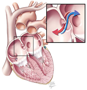
The first connection present in newborn hearts is the foramen ovale or atrial septal defect (ASD), an opening in the atrial septum between the two atria. Here is a diagram of the location of the foramen ovale:


(Image taken from: http://www.clevelandclinic.org)

The foramen ovale is used during fetal circulation to speed up the circuit time of blood through the heart. Fetuses don’t use their lungs because they receive oxygen rich blood from the mother via the placenta through the umbilical cord. Blood can therefore be directed straight from the right atria to the left atria without a need to travel through the right ventricle and pulmonary artery.

There is a similar hole in the ventricles referred to as the Ventricular Septal Defect (VSD), which joins the right and left ventricles through a patent hole in the ventricular septum. Just as in the foramen ovale, the VSD helps to mix oxygenated blood into the predominantly deoxygenated blood in the right ventricle and aorta.

Normally, the foramen ovale (and VSD) closes at birth due to increased blood pressure on the left side of the heart. The baby as well as the doctors can take advantage of the foramen ovale to prolong life without the need for major surgery. In some cases where the foramen ovale has already closed, minimally invasive catheterization surgery can be performed to make it patent using a small inflatable balloon similar to balloon angioplasty; The procedure is called a balloon septostomy.

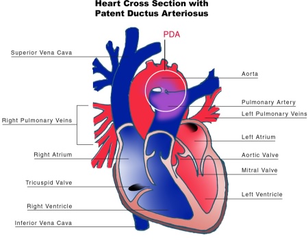
The third life-saving connection in newborns with great vessel transposition is a patent ductus arteriosus, a blood vessel that runs between the aorta and pulmonary artery. Here is a diagram of a patent ductus arteriosus:


As is the case with the foramen ovale, the ductus arteriosus begins to close shortly after the first breath. Generally, the ductus arteriosus completely closes four to ten days following birth. A small connection between the aortic branch and the left pulmonary artery remains after stenosis of the ductus arteriosus and is called the ligamentum arteriosum. Doctors can delay the stenosis of the ductus arteriosus by administering drugs such as Prostaglandin (Reference).

These three connections aside, no patient can sustain life with transposition of the great vessels because even with patent foramen ovale and ductus arteriosus there isn’t an adequate supply of oxygen to the tissue to maintain its viability, especially considering the added strain of the rapidly developing body.

There are two common surgical procedures used to correct for this malformation of blood vessels:
  • Mustard (or Senning) Operation – blood flow is corrected by transposing the pulmonary veins with the systemic veins.
  • Fontain Operation – also referred as the Arterial Switch Operation (ASO) – as the name implies this surgical procedure corrects the blood flow by de-transposing the great arteries of the heart.
Choosing the correct surgical procedure can be difficult because there are drawbacks with each. In the Mustard Operation, the procedure is much simpler surgically because veins are much easier to work with. The problem with the Mustard operation is that since the great vessels are still transposed the right side of the heart does the work of the left and vice-versa.

This becomes a problem when considering that each side of the heart performs a different job. The right side is intended to serve the lungs taking in deoxygenated blood from the systemic blood flow and reoxygenating it via the pulmonary arteries. There is considerable less resistance to flow in the right side of the heart and therefore the muscle is weaker than the left.

The left side has a more mechanically stressful job since it is required to push the blood throughout the body. From the start, each side of the heart is designed for different jobs and this means that ultimately the right side of the heart in a patient who has undergone a Mustard operation will fail under the intense stress of the muscle. These patients, whose hearts generally last for 20-30 years, will eventually need a transplant.

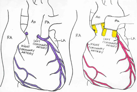
The Fontain or Arterial Switch procedure is more difficult surgically because the surgeon has to separate two major arteries (the aorta and pulmonary artery) from the ventricles and reattach them to the correct ventricle while also moving the coronary arteries, which are significantly smaller (1-2mm in an infant) and very important to proper heart growth and function. The pictures below show the patient’s heart pre- and post-operative. The X denotes the location of surgical stitches.


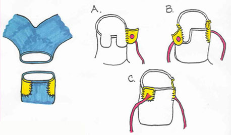
Normally the coronary arteries originate from the ascending aorta immediately distal from the aortic valve. When a surgeon is performing an Arterial Switch Operation (ASO), they must move the coronary arteries from the right side to the left so that the heart muscle receives oxygen-rich blood. To protect the coronary arteries, the surgeon removes a button of tissue surrounding the coronaries to aid in their attachment to the aorta. Here are two diagrams of the changes that are made during surgery:


Essentially, the surgeon excises the aorta and pulmonary arteries just above the valves and frees the coronaries (with additional muscle around it). Next, the surgeon sutures each coronary into place on the left side with fine precision (this can be seen in steps A-C in the first image above). The aorta is then moved into the correct position on the left side of the heart and sutured, above where the coronary arteries were sutured. Next, the two holes in the right side of the heart from the excision of the coronary arteries are patched with pieces of pericardium and the pulmonary artery is attached to the right side (as shown in the left of the first image above). Finally, the foramen ovale and ventricular septal defect are closed and the patent ductus arteriosus is tied off if open.

The surgery is complete but post-operative status is closely watched as complications such as bleeding, and/or myocardial infarction can occur. The patient who I saw this week was scheduled for routine scans to evaluate the condition of his heart. He had undergone a Fontain procedure as a baby and it is common for the patient’s physician to keep a close watch on the health of his heart.

(PAUSE for a quick breath) I guess I couldn’t get it all out in one breath. I hope that you enjoyed this case, I found it very interesting. Stay tuned for my next novel-length post coming soon!