This is my report for the first of six weeks in the Cornell Weill Medical College Summer Immersion program. My advisor,
Dr. Prince, has a dual appointment at Cornell University and Columbia University and is also the Chief of MRI at New York Hospital. Possibly his most noticeable contribution to science and the medical field is the development of contrast-enhanced Magnetic Resonance Angiography (MRA), which will be the subject of my post.
It would be very time consuming to first introduce Magnetic Resonance Imaging (MRI) and then explain contrast-enhanced MRA so I'll leave that to the experts.
If the sites above aren't enough to explain MRI then you'd be better off with a book on MRI. I would suggest Dr. Prince's book due out in August 2006 (
MRI From Picture to Proton). It may be a little expensive but it is by far the best reference on MRI and is the equivalent to the Dummy's Guide series of books.
Abbreviated Introduction to MRI
MRI as you should know by now stands for Magnetic Resonance Imaging. Now you might be asking yourself what magnetic resonance is and how can it be imaged. Lets break down the word MR:
Resonance is a condition in which the frequency of an external force matches the natural frequency of the object it is acting upon and
Magnetic refers to the presence of a magnetic field which induces the resonance. You might need to brush up on some E&M Physics to fully understand the concept of resonance. This magnetic resonance applies to certain atomic nuclei in your body such as
1H and
13C. For more information on nuclear magnetic resonance
read this.
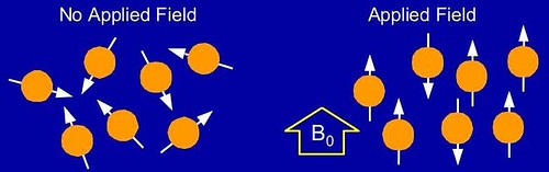
Nuclei that exhibit magnetic resonance are often referred to as nuclear spins and are normally oriented randomly. When these nuclei are placed in a strong magnetic field, they align with the applied field in their equilibrium state. The image below is a good example of this.
The MRI machine is a giant magnet that causes all of the
1H nuclei in your body to align along the applied field. Essentially, an image is acquired by applying a slightly different and smaller magnetic field to your nuclei that perturbs the alignment and determining how long it takes the nuclei to relax back to their original magnetization along B
0. Of course there are many variations on this principle that allow for the large array of different images that can be taken in MRI machine.
Contrast-Enhanced MRAMRA or Magnetic Resonance Angiography is a specific method of MRI that is utilized for imaging blood vessels. MRA is very useful for diagnosing problems with a patient's circulation, in particular vessel stenosis and/or aneurysm. This is an example of an MRA:

MRA is generally used to evaluate arteries in neck/brain, the thoracic and abdominal aorta, the renal arteries, and the legs (also referred to as run-off). A majority of the cases that I have seen in my first week have been patients with stenoses of the renal arteries and multiple stenoses of arteries in the leg (internal/external iliac, superficial femoral artery (SFA), popliteal, anterior tibial, peroneal, etc.) Problems with the vasculature are enhanced by Contrast-Enhanced MRA, which uses a contrast agent that accentuates the blood vessels - in particular the arteries - because of the paramagnetism of the contrast agent.
The most common and widely used contrast agent is Gadolinium (Gd); a rare earth element in the transition group IIIb of the periodic table. Gadolinium is abundant throughout the Earth's crust making it widely available but not necessarily cheap. The electron shell structure (eight unpaired electrons in the outermost shell) of Gd is what imparts its special paramagnetic properties. The paramagnetism of Gd in the body enhances the signal and thus intensity of the image depending where it is found in the body.
T1, the time it takes for longitudinal relaxation of the nuclear spin to return to equilibrium is affected by Gd as predicted by the following equation:
1/T1 = 1/1200 + (R1 * [Gd]) Eq. 1
R
1 is the T
1 value for the Gadolinium chelate, [Gd] is the concentration of gadolinium in the blood, and 1200 is the T
1 value for blood (in msec) without gadolinium. Gadolinium by itself is toxic and can cause heavy metal poisoning, but is safe for use in humans when bound to a chelator. The chelator does not affect the paramagnetism of gadlinium.
Contrast-Enhanced MRA uses a small (30-60 mL) bolus injection of Gd which is then followed by acquiring a series of images that track the flow of Gd through the body. The technician operating the machine will acquire a series of images at different times in the same location. This allows for a time-lapsed movie of the contrast agent flowing through the vasculature. However, timing of images and location is vital to ensuring that adequate detail is obtained from the Contrast MRA. For more information on contrast-enhanced MRA read
this article. A sample of a MRA that has been imaged over time to show the change is contrast in the arteries can be found
here.
One of the aspects of contrast-enhanced imaging is in some procedures the patient is required to hold their breath in order to reduce motion artifacts in subsequent images. The biggest problem with breath-holding is that the process of inhale-exhale-hold takes a significant amount of time that makes the scanning procedure long and uncomfortable for the patient and technician. All breath-hold imaging is done during expiration because it is easier to hold one's breath when the lungs are deflated.
The problem is that people can hold their breath longer during inspiration but the motion in images caused by holding your breath during inspiration is too great to make imaging effective. The MRI technician observes the respiratory phases through a small elastic belt located on their chest just below the xiphoid process
(small cartilaginous protrusion from bottom of rib cage).
One school of thought is that if people were enabled to hold their breath for longer the imaging procedure could be sped up and time and money saved in the process. Increasing breath-holding can be accomplished several ways, the most straightforward way is to administer the patient a hyperoxic gas. Hyperoxic gas is simply air that has an excess of oxygen; typical room air has ~20% Oxygen but by increasing the O2 level to 40%, studies have shown that people can hold their breath longer than under normoxic conditions.
My project for this summer will be to design, develop, and test a device that is MR compatible and will administer hyperoxic gas to the patient. One of the biggest concerns is making the device simple enough that it can be setup in a matter of seconds.
MRA is certainly not as simple as it looks and there are a lot of variations in the procedure that I am not yet familiar with. As I learn and work on my project, I will be posting about my progress here.